- Solid State Laser
- Semiconductor Laser
- DPSS laser
- Gas Laser
- Fiber Laser
- Fiber Coupled Laser(MM)
- Fiber Coupled Laser(SM)
- Ultrafast laser
- Femtosecond Laser
- Picosecond Laser
- Pulsed Laser
- Q-switched Laser
- Raman Laser
- Narrow Linewidth Fiber Laser
- Wavelength Tunable Laser
- Laser Diode
- Laser Module
- Pigtailed Laser
- DFB Laser
- DBR Laser
- SLED Laser
- VCSEL Laser
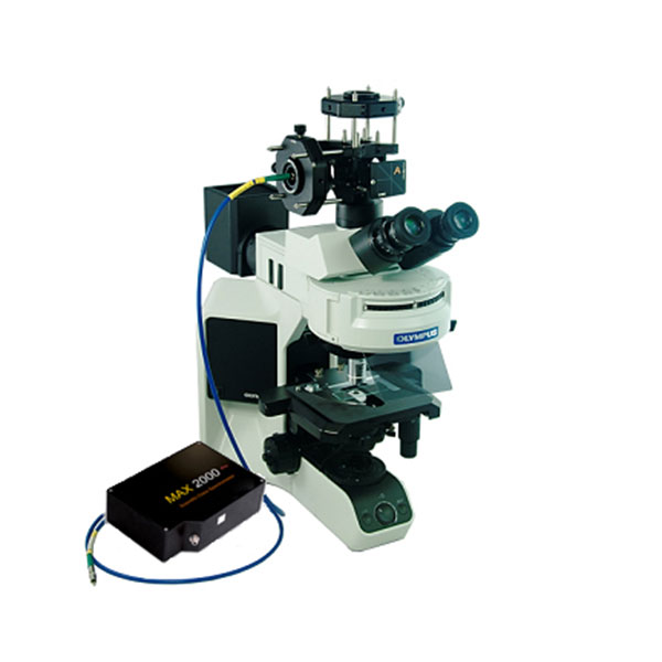
Microscopic Spectrometer Microscopic Angle Resolution Microscopic Fluorescence Microscope Spectrum
Product ID: 6121
$22,707 – $45,400
Microscopic Spectrometer Microscopic Angle Resolution Microscopic Fluorescence Microscope Spectrum
Microspectroscopy system:
Using the world’s best Semrock filter set, creatively integrate the excitation light, fluorescence and filter into one probe. At the same time, with the micro-area probe coupling module, the spatial resolution of fluorescence spectrum measurement can be increased to 5μm. The microscopic spectroscopy system, as the name implies, is the combination of a microscope system and a spectrometer system. It has both the function of microscope imaging and the function of spectrum analysis. The system can realize the fluorescence spectrum, reflection spectrum, transmission spectrum, Raman spectrum and other spectral analysis of micron-level samples, and is widely used in the fields of materials, biotechnology, mineral analysis, micro-nano optics and other fields.
System Model: MS-Motic-XXX / MS-Olympus-XXX/ MS-Leica-XXX (Choose)
Microscope brand: Motic / Olympus/ Leica (Choose)
XXX in the model number stands for fiber spectrometer:
Fiber spectrometer: EQ2000 / EQ4000 / PC2000 / … (Customized)
Microscopic spectrometer system:
.jpg)
Structure and composition:
The microspectroscopy system can be divided into three modules: lighting module, spectrum receiving module and imaging module.
1. Illumination module
The lighting modules of the microspectroscopy system are generally divided into two types: Kohler lighting and confocal lighting.
a) The light source of Kohler illumination is generally the halogen lamp that comes with the microscope, and the halogen filament is imaged on the back focal plane of the objective lens through the lens group. In this way, the object can obtain brighter and uniform full-field illumination; the principle diagram can be seen in Figure 1 .
.jpg)
b) Confocal illumination is to introduce the illumination light source (such as laser, xenon lamp, etc.) into the microscope spectroscopy system through the optical fiber. The output end surface of the optical fiber is imaged on the object surface through the optical system, that is, the incident end surface and the object surface are conjugated to achieve fixed-point illumination or excitation .
2. Spectral receiving module
The module is composed of an optical fiber and a miniature spectrometer, in which the optical fiber receiving optical path is confocal reception, that is, the receiving surface and the object surface are conjugate surfaces to realize fixed-point spectrum reception. One end of the receiving fiber is connected to the optical path of the microscope, and the other end is connected to a miniature spectrometer, so as to obtain the spectral information in the microscopic area of the object.
3. Imaging module
The module is a CCD camera. On the basis of a microscope, the CCD/CMOS camera is placed on the conjugate surface of the object surface. While measuring the spectrum, it can realize the real-time acquisition of object images, that is, conjugate imaging.
.jpg)
System Features:
1) Easy to operate: The microspectroscopy system is improved and optimized based on the optical path of the microscope, and a spectrum measurement module is added. The measurement procedure can be divided into two steps, one is to find the object under the microscope to make the object appear a clear image under the eyepiece, and the other is to collect the spectrum through the micro-spectrometer software.
2) The object is small and the area is optional: Using the principle of confocal, the receiving fiber can only receive the area where the fiber end face is imaged on the object surface, so as to realize the spectrum collection in a small area. The spatial resolution of the acquisition area can generally be obtained by dividing the core diameter of the receiving fiber by the magnification of the objective lens. Through a specially customized optical fiber, a ring can be formed around the collection area to realize the area selection and positioning of small objects.
3) Strong measurement capability: It has spectral measurement functions that traditional microscopes do not have. Traditional microscopes can only provide image acquisition, so that the topography of the object can be analyzed, and the spectrum information of the object cannot be obtained. The microscopic spectrum measurement system, in addition to the function of collecting object images, can also collect and analyze the spectrum of the object in different regions, and further understand the structure and characteristics of the object.
4) Multiple extended functions: Based on a commercial microscope, through the design and coupling of the optical path switcher, the transmission and reflection, fluorescence, and Raman spectroscopy measurements under the microscope can be added to meet various scientific research needs to the greatest extent.
Typical microscopic spectrum measurement
1) Microscopic reflection spectrum measurement: usually use the halogen lamp that comes with the microscope as the illuminating light source, illuminate the object through the upper reflection light path in the microscope (Kohler illumination), and enter the receiving fiber after being reflected by the object. Spectroscopy and analysis of reflected light.
2) Microscopic transmission spectrum measurement: usually use the halogen lamp that comes with the microscope as the light source, illuminate the object through the transmission light path under the microscope, and the light reaches the receiving fiber after passing through the object, and the micro-spectrometer is used to collect the spectrum of the received transmitted light And analysis.
3) Microscopic fluorescence spectrum measurement: The external laser light source is coupled into the microscope system through the optical path switcher through the optical fiber or fluorescent probe, and is focused on the object surface to realize the fluorescence excitation of the object. Then, by filtering the light returned from the excited point (filtering out the excitation laser), the light entering the receiving fiber only retains the required fluorescence information, and the micro-spectrometer is used to spectrum and analyze the received fluorescence.
4) Microscopic Raman spectroscopy measurement: The external laser light source (wavelength is 532nm or 785nm) is coupled into the microscope system through the optical path switcher through the Raman probe, and focused on the object surface to realize the Raman excitation of the object. Then, by filtering the light returned from the excited point (filtering out the excitation laser), the light entering the receiving fiber only retains the required Raman and fluorescence information, and the micro-spectrometer is used to measure the received Raman light and fluorescence. Collection and analysis.
MS-Microscopic Spectrum Analysis System:
.jpg)
| Model |
|---|
